
Axon-Dendrite Classification
Multi-cue strategy for distinguishing axons from dendrites in EM — morphology, organelles, synaptic role, continuity, and confidence scoring. Full instructor script with worked examples.

Overview
Misclassifying a process as axon when it is actually a dendrite (or vice versa) is one of the most consequential annotation errors in connectomics. It corrupts the directed graph: an axon→dendrite synapse becomes nonsensical if the “dendrite” is actually an axon. Cell-type identification, input/output analysis, and circuit motif detection all depend on correct process classification. This document provides a systematic, multi-cue approach to axon-dendrite discrimination that can be taught to new annotators and used as a calibration reference for experienced teams.
Instructor script: the classification challenge
Why is this hard?
In textbook diagrams, axons and dendrites look obviously different — axons are thin and smooth, dendrites are thick and spiny. In real EM data, the distinction is often far less clear:
- Thin dendrites in distal arbors can be <200 nm diameter, rivaling the caliber of unmyelinated axons
- En passant boutons (axonal swellings with vesicles) can resemble dendritic spines at certain angles
- Truncated processes at volume boundaries lack the full morphological context needed for confident classification
- Glial processes can be confused with both axons and dendrites (see glia-recognition.md)
- Developing or immature tissue has less distinct morphological signatures
The key principle: no single cue is reliable alone; classification requires convergent evidence from multiple independent features.
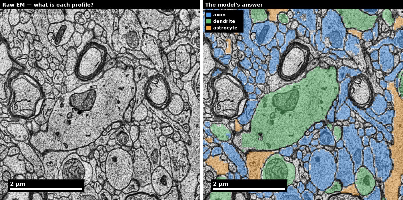
scripts/render_em_figures.py.
The four-pass classification protocol
Pass 1: Morphology
Evaluate the process shape and branching pattern:
| Feature | Axon-like | Dendrite-like |
|---|---|---|
| Caliber | Thin, relatively uniform (0.1-1.0 μm) | Thicker proximally (5-10 μm), tapering distally |
| Branching | Frequent, often at acute angles | Fewer branches, often at wider angles |
| Spines | Absent (except rare axonal filopodia) | Present on most excitatory dendrites |
| Surface texture | Smooth | Irregular (spines, varicosities) |
| Tapering | Minimal (uniform caliber over long distances) | Progressive (proximal → distal gradient) |
| Tortuosity | Often more tortuous | Generally smoother trajectory |
Caveat: Inhibitory neuron dendrites are often smooth (aspiny), so spine absence does not automatically mean “axon.”
Pass 2: Organelles
Evaluate the intracellular contents:
| Feature | Axon-like | Dendrite-like |
|---|---|---|
| Synaptic vesicles | Present (especially at boutons): 40-50 nm clear vesicles clustered near membrane | Absent (or only in rare dendritic release sites) |
| Microtubules | Uniformly plus-end-out (vertebrates); often bundled | Mixed polarity; more evenly distributed |
| Ribosomes/RER | Absent (beyond the axon initial segment) | Present (polyribosomes at spine bases, RER in shaft) |
| Mitochondria | Small, elongated (0.5-2 μm) | Larger, more branched, often clustered near active spines |
| Smooth ER | Thin tubule running along the axon | Network; forms spine apparatus in spines |
| Dense-core vesicles | Present in some axon types (80-120 nm) | Rare |
Key cue: Vesicle clusters are the strongest single indicator of axonal identity. If you see a cluster of ~40 nm clear vesicles near a membrane, the process is almost certainly an axon (at that location, it is acting as a presynaptic terminal).
Pass 3: Synaptic role
Evaluate how the process participates in synapses:
| Role | Axon | Dendrite |
|---|---|---|
| Presynaptic | Yes — forms output synapses | Rare (dendro-dendritic synapses exist but are uncommon in cortex) |
| Postsynaptic | Rare (axo-axonic synapses onto AIS from chandelier cells) | Yes — receives input synapses |
| Spine-bearing | No | Yes (excitatory neurons) |
Decision rule: If the process is consistently presynaptic (vesicles on its side, PSD on the partner’s side), it is an axon. If consistently postsynaptic, it is a dendrite. If it shows both roles at different locations, investigate carefully — it may be a merge error joining an axon and dendrite.
Pass 4: Continuity
Verify the interpretation by following the process through additional sections:
- Trace proximally: Does the process eventually connect to a soma? If so, how? Axons typically emerge from the axon hillock (one per neuron). Dendrites emerge as multiple thick trunks.
- Check consistency: Does the classification remain stable along the process? A sudden change from “axon-like” to “dendrite-like” features suggests a merge error.
- Branching pattern: Axonal arbors tend to have more terminal branches. Dendritic trees tend to have a recognizable morphological type (apical, basal, stellate).
Edge cases and how to handle them
Thin, aspiny dendrites of inhibitory neurons
Problem: Inhibitory interneuron dendrites (e.g., parvalbumin+ basket cells) are often smooth (no spines), thin (0.3-1.0 μm), and extend long distances — features that overlap heavily with axons.
Solution: Look for (a) postsynaptic role — these dendrites receive input synapses on their shaft, (b) ribosomes/RER in the cytoplasm (absent in axons), (c) microtubule polarity if assessable, (d) trace to the soma and check cell type.
En passant boutons vs spines
Problem: An axonal en passant bouton (swelling along the axon shaft containing vesicles) can resemble a spine head when viewed in a single section.
Solution: (a) Check for vesicles — boutons contain vesicle clusters, spine heads typically do not. (b) Check PSD position — in a bouton, the PSD is on the partner’s membrane; in a spine, the PSD is on the spine head membrane. (c) Follow the process through z — boutons are continuous with the axon shaft; spines connect to a larger dendrite via a narrow neck.
Truncated processes at volume boundary
Problem: A process is cut by the edge of the imaged volume. You can see only the last few micrometers, not enough to trace to a soma or evaluate the full branching pattern.
Solution: Use the available local cues (organelles, vesicles, synaptic role) and assign a confidence level. Mark as provisional if evidence is insufficient. Do not force a classification — uncertain labels are more valuable than incorrect ones.
Myelinated processes
Problem: Myelin wrapping obscures the cytoplasm, making organelle-based cues less accessible.
Solution: Myelin itself is a strong axon indicator (dendrites are never myelinated in mammals). For the myelinated segment, classify as “myelinated axon” with high confidence. At nodes of Ranvier (gaps in myelin), cytoplasmic features become accessible for additional confirmation.
Worked example: classifying an ambiguous process
Scenario: A process ~300 nm in diameter runs through 15 consecutive sections. It contains a few microtubules, no obvious vesicle clusters, and no spines. It receives one synapse (PSD on its side) and forms no presynaptic contacts in the visible region.
Pass 1 (Morphology): 300 nm is ambiguous — could be a thin dendrite or a thick unmyelinated axon. No spines. Smooth surface. Verdict: inconclusive.
Pass 2 (Organelles): A few microtubules (3-5), one small mitochondrion. No vesicles. No ribosomes visible. Verdict: slight lean toward axon (no ribosomes), but insufficient evidence.
Pass 3 (Synaptic role): One synapse where this process is postsynaptic. Verdict: lean toward dendrite. Axons rarely receive synapses (exception: axo-axonic chandelier cell inputs onto the AIS, but this is not near a soma).
Pass 4 (Continuity): Trace 10 sections proximally. The process thickens slightly (350 nm) and a ribosome cluster becomes visible near a branch point. Two more input synapses appear on the shaft.
Final classification: Dendrite (likely inhibitory interneuron, given smooth morphology). Confidence: medium. Key evidence: postsynaptic role (3 input synapses), ribosomes detected in proximal shaft. Uncertainty: no spine evidence (consistent with inhibitory neuron), limited tracing distance.
Inter-annotator calibration protocol
To maintain consistent classification across a team:
- Weekly calibration sessions: Review 10-20 ambiguous cases as a group. Discuss cue evidence and arrive at consensus.
- Shared edge-case library: Maintain a documented set of difficult examples with agreed-upon classifications and reasoning.
- Confusion matrix tracking: Periodically test annotators against an adjudicated truth set. Track axon/dendrite classification accuracy per annotator.
- Threshold agreement: Set team-wide rules for minimum evidence required before committing a classification (e.g., “at least two independent cue types must agree”).
How reliable is each cue?
The protocol above lists cues; it does not say which ones fail, and that is the information you need to work under uncertainty. Roughly, in decreasing order of reliability:
| Cue | Reliability | Fails when |
|---|---|---|
| Polyribosomes / rough ER present | Very high for dendrite. Axons lack them | The section misses them; a thin dendrite may show none in any single plane |
| Vesicle-filled varicosity with an active zone | Very high for axon | The bouton is out of plane; a dendrite receiving many inputs can look busy at low magnification |
| Postsynaptic densities on the process itself | High for dendrite | Axo-axonic synapses exist, and are common on the AIS specifically |
| Myelination | Very high for axon | Only a minority of axons are myelinated, so absence proves nothing |
| Microtubule arrangement | Moderate | Both compartments contain microtubules; the difference is spacing and fasciculation, which needs a clean cross-section |
| Spines | High for dendrite | Aspiny and sparsely spiny dendrites are common — most inhibitory neurons |
| Calibre | Low on its own | Thin dendrites and thick axons both exist. Unit 06 §1 gives 80-300 nm for unmyelinated neurites and 50-200 nm for spine necks — overlapping ranges |
| Branching angle | Low | Suggestive at best, and strongly affected by section angle |
The rule the units enforce: two independent cue families before a confident call. Independence is the operative word. Calibre and branching angle are both geometric and both fail on a tangential cut, so they are one family, not two. Organelle content and synaptic role are genuinely independent of geometry, and that is why they carry the call.
Attaching a confidence tier
Use the same three-bin vocabulary the rest of the site uses, so a call travels with its evidence:
- Bin A — two independent cue families agree, both clearly visible. Record the call and which cues you used.
- Bin B — one strong cue, or two weak agreeing ones. Record the call and what would change your mind.
- Bin C — cues conflict, or the section is uninformative. Record “uncertain”. This is a real answer, and a dataset where nobody ever writes it is a dataset where people are guessing.
A tier is only useful if it predicts accuracy. Unit 06’s rubric asks that high-confidence calls be right at least 90% of the time while a non-trivial share stay uncertain; if your Bin A accuracy matches your overall accuracy, the tiers are decoration.
Species and tissue caveats
Everything above is calibrated on mammalian cortical neuropil. It does not transfer unchanged:
- In Drosophila, most neurites are unmyelinated, many neurons are unipolar, and the same process can be pre- and postsynaptic along its length — the axon/dendrite dichotomy is often the wrong frame entirely.
- In C. elegans, processes are thin enough that organelle cues are frequently absent from a given section.
- In human tissue (H01 and comparable surgical material), fixation quality varies with depth from the resection surface, and the first cue to degrade is membrane contrast — which is the one the automated segmentation depends on.
Before applying this protocol to a new dataset, classify twenty processes you can verify and check that the cues behave as described. If they do not, it is the protocol that needs adjusting, not the tissue.
Common misconceptions
| Misconception | Reality | How to verify |
|---|---|---|
| “Thin process = axon” | Thin aspiny dendrites are common in inhibitory neurons | Check synaptic role and ribosomes |
| “No spines = axon” | ~20-30% of cortical neurons are inhibitory with smooth dendrites | Trace to soma; check cell type |
| “Vesicles = always axon terminal” | Dense-core vesicles can be in transit through dendrites; some dendrites release transmitter | Check for clustered clear vesicles specifically at membrane appositions |
| “One synapse proves the classification” | A single synapse can be misdetected or ambiguous | Require multiple synapses or corroborating organelle evidence |
References
- Baas PW et al. (1988) “Polarity orientation of microtubules in hippocampal neurons: uniformity in the axon and nonuniformity in the dendrite.” PNAS 85(21):8335-8339.
- Harris KM, Weinberg RJ (2012) “Ultrastructure of synapses in the mammalian brain.” Cold Spring Harbor Perspectives in Biology 4(5):a005587.
- Kasthuri N et al. (2015) “Saturated reconstruction of a volume of neocortex.” Cell 162(3):648-661.
- Peters A, Palay SL, Webster HdeF (1991) The Fine Structure of the Nervous System. 3rd ed. Oxford University Press.
- Schneider-Mizell CM et al. (2016) “Quantitative neuroanatomy for connectomics in Drosophila.” eLife 5:e12059.