Recognizing astrocytes, oligodendrocytes, and microglia in EM, why glia-neuron boundary errors corrupt neuronal connectivity, and a drill-based protocol for getting them right.
Key community resources for this unit:
Savage, Picard, González-Ibáñez & Tremblay (2018)
— "A Brief History of Microglial Ultrastructure: Distinctive Features, Phenotypes, and Functions Discovered Over the Past 60 Years by Electron Microscopy," Frontiers in Immunology 9:803 Review specifically on EM identification of microglia.
Chiappini et al. (2025)
— "Ultrastructure of astrocytes using volume electron microscopy: A scoping review," The Journal of Physiology 604(4):1498–1517 Current, directly on-topic review of astrocyte identification via volume EM.
Peters, Palay & Webster, The Fine Structure of the Nervous System
Includes dedicated chapters on oligodendrocyte, astrocyte, and microglial ultrastructure — the oligodendrocyte anchor here, since no single modern review paper on oligodendrocyte EM identification was as canonical as the microglia/astrocyte reviews above (worth a closer look if you want a dedicated paper instead).
Before you start
Time
Self-study ~2.5 h: about 90 min of reading plus the 60 min discrimination drill. Taught: a 90 min session, per the lecture plan. Deck: the unit’s slide deck is scoped to 60 min and does not follow the plan slide for slide.
Prerequisites
Units 05–06
You need
A public EM volume; the Unit 05 organelle table
You finish with
A glia recognition checklist and a measured discrimination score by cell class
The claim this unit has to earn: glia are not background, and correcting glia is
not tidying up. Glia and their processes occupy roughly 20–40% of cortical tissue
volume, and astrocytic processes specifically form a fine meshwork that interleaves
with every neurite in the neuropil. That geometry is exactly what makes them the most
frequent partner in a merge error.
What you’ll be able to do
Identify astrocytes, oligodendrocytes, and microglia from soma and process features.
Discriminate a fine astrocytic process from a thin neurite using cues that survive poor staining.
Explain, with a concrete mechanism, how a single glia-neuron merge corrupts a connectivity measurement.
Prioritize glia corrections against other proofreading work on impact rather than count.
Run a discrimination drill and interpret the resulting confusion matrix.
1. Why a glia merge is expensive
Take one merge: a fine astrocytic process is fused with a nearby dendrite.
What happens to the neuron:
It gains a branch that does not exist. Its total dendritic length, branch count, and
arbor extent are all wrong.
The astrocytic process wanders through neuropil the real dendrite never visits.
Synapse detections along that path get attributed to this neuron — so the neuron
gains inputs from cells it has never contacted.
Because astrocytic processes ensheathe synapses, the merged path runs directly
past a large number of synapses. The false-input yield per unit length of an
astrocytic merge is unusually high.
What happens to the astrocyte: it loses territory, so any astrocyte morphometry —
domain volume, synapse coverage, vascular contact — is also wrong.
What happens to your analysis: the false inputs are not random. They are spatially
local, which means they preferentially connect the neuron to its neighbors. A
motif analysis will see enhanced local clustering; a distance-dependence analysis will
see inflated short-range connectivity. Again the bias points toward an interesting
result.
Consequence for triage. Glia-neuron merges should be ranked above many
neuron-neuron splits in a proofreading queue, even though a split is more visually
obvious. The Unit 08 rule — prioritize by effect on the endpoint metric, not by
conspicuousness — has its clearest application here.
Check yourself
A pyramidal cell in your analysis set turns out to be merged with a fine
astrocytic process. Before the fix it had noticeably more inputs, and a strong local
clustering coefficient. Which of your results move when the merge is removed, and in
which direction?
Everything that counted the false inputs moves toward the less interesting answer.
Input count and dendritic length fall. The merged path was a branch that does not
exist, and it ran past synapses the real dendrite never contacted.
Local clustering and short-range connectivity fall. The false inputs were not
random: they came from the cell’s spatial neighbours, because that is where the
astrocytic process ran. A merge like this inflates exactly the statistics that make
a circuit look locally wired.
The astrocyte’s own morphometry — domain volume, synapse coverage — changes too,
because it gets its territory back. If the project makes any glial claim, that
number was also wrong.
What does not change is how the neuron looked before the fix. That is the problem:
nothing about the merged object announced the error.
Generalizable principle: when an error’s false positives are spatially local, the
bias is toward local structure. Ask where the error’s extra material came from before
deciding it is noise.
2. Recognition: the three classes
Each class has one near-diagnostic feature. Learn those three first; everything else
is corroboration.
Astrocyte — diagnostic feature: glycogen granules
Nucleus
Pale, euchromatic, often irregular in contour
Cytoplasm
Pale and “watery”; few organelles; bundles of intermediate filaments (GFAP) ~10 nm
Glycogen granules
20–30 nm, very dark, in clusters. Neurons do not contain them.
Soma size
~8–10 µm, smaller than most neuronal somata
Processes
Irregular, sheet-like, fill the gaps between neuronal elements rather than running as cylinders
Perisynaptic processes
Extremely thin (< 100 nm), wrapping synapses — the hardest structures in the volume to segment
Synaptic vesicles, PSDs, and (largely) microtubules
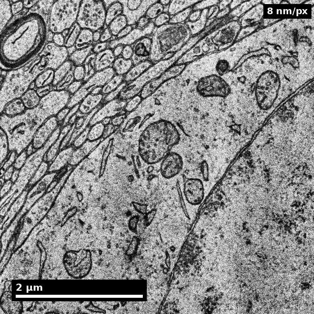
Astrocytic territory in human cortex, labelled astrocyte by H01's subcompartment model rather than picked by eye. Look for the pale, organelle-poor cytoplasm and the outline that takes whatever shape its neighbours leave. Then look for the diagnostic feature in the table above and notice how hard it is: glycogen granules are 20–30 nm, which is under four pixels at this rendering. Not seeing them here is the weak evidence the check-yourself in this section warns about.H01 human cortex, Lichtman Lab (Harvard) & Connectomics at Google, CC BY 4.0. Shapson-Coe et al., Science 384, eadk4858 (2024). Rendered by scripts/render_em_figures.py.
Oligodendrocyte — diagnostic feature: the darkest nucleus in the field
Nucleus
Small, round, extremely electron-dense heterochromatin. Distinctly darker than neuronal or astrocytic nuclei — usually identifiable at a glance and at low magnification
Cytoplasm
Dense, abundant rough ER and ribosomes, prominent Golgi, microtubules present
Soma size
~6–8 µm
Processes
Connect the soma to myelin sheaths; each cell myelinates on the order of 20–60 axonal segments
Myelin relationship
Inner and outer tongues contain oligodendrocyte cytoplasm; paranodal loops at nodes of Ranvier
The practical difficulty is not the mature oligodendrocyte — it is the
oligodendrocyte precursor cell (OPC/NG2 cell), which has a paler nucleus and can
resemble a small neuron or an astrocyte. If a cell looks “sort of oligodendrocyte but
the nucleus is not dark enough”, OPC is the leading hypothesis and the correct action
is usually to flag rather than force.
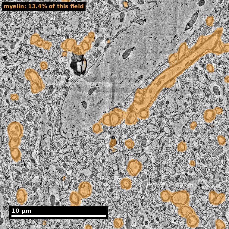
The oligodendrocyte's footprint in neuropil: every myelinated axon in this grey-matter field, highlighted by H01's myelin mask, which covers 13.4% of the field. Each sheath is oligodendrocyte membrane, made by a cell whose soma is not in view. What step 5 of the protocol keys on — the inner and outer tongues of oligodendrocyte cytoplasm at the sheath's edges — is not resolvable at this magnification; the mask tells you where to zoom in, not what you would find.H01 human cortex, Lichtman Lab (Harvard) & Connectomics at Google, CC BY 4.0. Shapson-Coe et al., Science 384, eadk4858 (2024). Rendered by scripts/render_em_figures.py.
Microglia — diagnostic feature: dense elongated nucleus plus lysosomal content
Nucleus
Dark, often elongated or bean-shaped, with heterochromatin clumped along the nuclear envelope
Cytoplasm
Dense, with characteristic long, narrow ER cisternae; lysosomes, phagosomes, and lipofuscin-like inclusions
Processes
Fine, highly irregular contours; frequently seen contacting synapses and vessels
State-dependence
Morphology changes substantially with activation state, which makes microglia the least stereotyped of the three
Astrocyte vs thin neurite: the discrimination that matters most
This is where most errors occur, so it gets its own table.
Feature
Astrocytic process
Thin neurite
Glycogen granules
Present
Absent
Synaptic participation
None (no vesicles, no PSD)
Vesicles (axon) or PSD (dendrite)
Microtubules
Absent or very rare
Usually present
Cross-sectional shape
Irregular, angular, sheet-like; fills residual space between other profiles
Roughly circular or elliptical; a coherent tube
Cytoplasm
Pale, few organelles
Denser, organelles visible
Intermediate filaments
Fine bundles (GFAP)
Absent (neurofilaments differ in distribution and context)
Trajectory across sections
Wanders, changes shape section to section
Maintains a continuous, traceable trajectory
The shape cue is underused and it is robust. Astrocytic processes are space
filling — their cross-section is whatever shape is left over after the neurites are
packed. Neurites are tubes — their cross-section is a shape in its own right. This
cue survives weak staining better than glycogen granules do, which makes it valuable
exactly in the regions where everything else fails.
Check yourself
A pale, irregular profile sits between three neurites. You see no glycogen
granules. Is it an astrocytic process?
Probably, but this is a medium-confidence call at best, and the reasoning matters
more than the answer.
Absence of glycogen granules is weak evidence here: granules are clustered and
sparse, so a given thin process may contain none in a given section even when it is
astrocytic. This is the Unit 06 lesson about over-reading absence.
Better evidence to seek, in order:
Shape and space-filling across several sections — does it change contour to
fill gaps, or does it hold a tube shape?
Synaptic participation — scroll for any vesicle cluster or PSD on it. Finding
one converts this to a neurite call immediately.
Continuity toward a soma or an endfoot — following it to a vessel endfoot is
close to definitive for astrocyte.
Microtubules at higher magnification.
If cues 1–4 do not resolve it, the correct output is uncertain, with a note that
the deciding cue was not resolvable. That is a useful annotation.
3. The identification protocol
1. Is this a soma (nucleus visible)?
   YES -> nucleus first:
      very dark, small, round      -> oligodendrocyte (check for myelin links)
      dark, elongated, peripheral
        heterochromatin + lysosomes-> microglia
      pale, irregular, glycogen    -> astrocyte
      pale, round, big nucleolus,
        Nissl/rER, dendrites       -> neuron
      pale but "not quite"         -> OPC candidate -> FLAG
   NO  -> continue to 2

2. Does the process participate in a synapse
   (vesicle cluster or PSD on it)?
   YES -> neurite. Go to Unit 06 for axon/dendrite.
   NO  -> continue

3. Glycogen granules present?
   YES -> astrocyte (high confidence)
   NO  -> continue (absence is weak evidence)

4. Cross-sectional character over 5-10 sections:
   space-filling, contour changes to fit gaps -> astrocyte (medium-high)
   coherent tube, stable contour              -> neurite (medium)

5. Context:
   wraps a capillary (endfoot)        -> astrocyte (high)
   inner/outer tongue of a myelin
     sheath, or paranodal loop        -> oligodendrocyte (high)
   irregular contour + lysosome-rich
     parent process                   -> microglia (medium)

6. Unresolved -> UNCERTAIN, with the missing cue named.
   Route to the glia review queue.
Worked example: a flattened profile against a capillary
Patch: a pale profile, a few hundred nanometers thick and about 2 µm long,
pressed flat against the wall of a capillary. The parent process leaves the
plane. The segmentation currently assigns it to a nearby dendrite — so this is
also a candidate merge, and the call decides an edit.
Step 1 — soma? No nucleus in view. Continue.
Step 2 — synaptic participation? Scroll several sections in each direction:
no vesicle cluster, no PSD anywhere on the profile. This step is not skippable
even when the vascular context is already shouting the answer. A perivascular
call made without ruling out synaptic participation is exactly how a dendrite
that happens to end near a vessel stays merged into an astrocyte — or the
reverse.
Step 3 — glycogen? None visible. Weak evidence: granules are clustered and
sparse, so one thin profile showing none decides nothing. Noted, not spent — the
same discipline as the §2 check-yourself.
Step 4 — cross-sectional character over 5–10 sections. The contour does not
hold a tube shape. It spreads and flattens to track the vessel wall, section
after section — space-filling behavior. Astrocyte-leaning, medium confidence on
its own.
Step 5 — context. A flattened expansion covering a vessel surface is the
endfoot configuration, and the protocol scores a capillary wrap as
high-confidence astrocyte. But one alternative has to be dispatched before
taking that: microglial processes also contact vessels, and this is where an
honest annotator hesitates. The unit’s own recovery rule supplies the test —
microglia require a nucleus or a lysosome-rich parent process, never a fragment.
Follow the profile away from the vessel: the parent is pale, organelle-poor,
with fine intermediate-filament bundles, and none of the dense, inclusion-rich
cytoplasm a microglial process trails back to. Microglia is dismissed for cause,
not by default.
Call: astrocytic endfoot; high confidence. The dendrite assignment is
therefore a glia–neuron merge, and per §1 it goes into the queue above more
conspicuous splits. Evidence chain: space-filling sheet geometry across sections
plus vessel-covering context — two independent cues — with synaptic
participation ruled out and the microglial alternative rejected on cytoplasmic
evidence. Steps 2 and 3 contributed only absences, and were weighted as
absences.
Transferable principle: context is the strongest cue in the glial protocol,
but it is only safe after the exclusion steps have run. Work the cheap
rule-outs first even when the context looks decisive — especially then, because
a decisive-looking context is what tempts you to skip them. Two further cases —
astrocyte versus thin dendrite, and OPC versus small neuron — are worked in
Glia recognition.
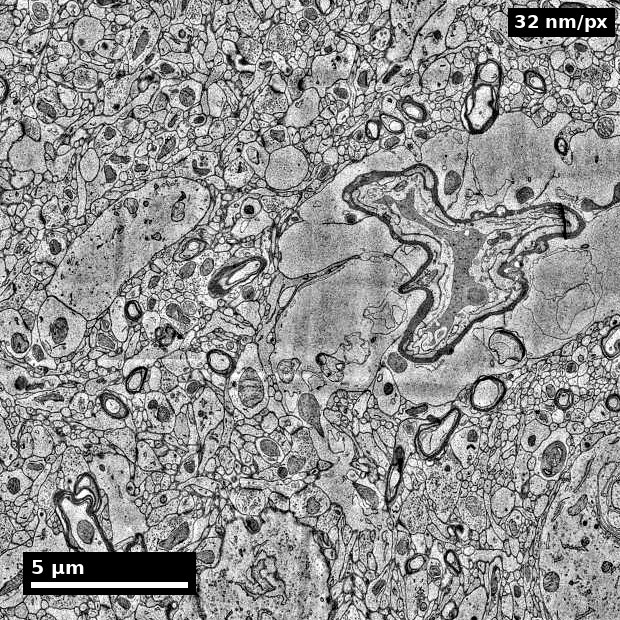
A cortical blood vessel, located by H01's own vessel mask rather than chosen by eye. This is the context step 5 of the protocol scores highest for astrocyte. Notice what the image does not tell you: at 32 nm per pixel, in one plane, you cannot say which of the thin profiles against the wall are endfeet. The worked example above reached its call only after the exclusion steps, across several sections.H01 human cortex, Lichtman Lab (Harvard) & Connectomics at Google, CC BY 4.0. Shapson-Coe et al., Science 384, eadk4858 (2024). Rendered by scripts/render_em_figures.py.
Check yourself
A soma has a round nucleus that is clearly darker than the neurons around it,
but noticeably paler than the oligodendrocyte two fields over. There is no myelin in
view. A trainee labels it "OPC", with a note: "not sure". Grade the call.
The call may be right; the note shows the label is being used for the wrong thing.
The appearance fits the protocol’s “pale but not quite” branch: an oligodendrocyte
precursor has a paler nucleus than a mature oligodendrocyte and can resemble a small
neuron or an astrocyte. So OPC candidate is a reasonable hypothesis, and the
protocol’s action for it is flag, not force.
What fails is the note. “OPC” and “uncertain” are different annotations with different
follow-ups — an OPC candidate goes to someone who can confirm a cell class, an
uncertain call goes to the review queue with the missing cue named. A trainee who
writes “OPC, not sure” has merged the two, and if that habit spreads, per-class
agreement for OPC stops meaning anything — and OPC agreement is usually the worst of
the four classes already (§5).
A better record: OPC candidate — nucleus intermediate in density; no myelin link found
in view; neuronal features (large nucleolus, Nissl, dendrites) not seen. That names
the evidence for and against, which is what lets a reviewer settle it.
Generalizable principle: a label is a claim, and “uncertain” is its own label. Never
let a class name stand in for a confidence level.
Visual training set
Work these against the identification protocol in §3, naming the step that decides each case. They are single planes and the decisive glial cue usually is not one: step 4 asks how a cross-section behaves over five to ten sections, and perisynaptic astrocytic sheets under 100 nm thick are the hardest structures in the volume. Use the panel as a reference for what the cues look like, and do the calling in a volume you can scroll through z.
RIV-GLIA S01: Orientation for glia proofreading. Set the stake before you start looking: glia occupy roughly 20–40% of cortical volume, and a glia-neuron merge does not merely add a branch — it drags a neuron’s arbor past synapses it never contacted, so the false-input yield per micrometer of merged path is unusually high.
RIV-GLIA S03: Astrocyte morphology in a synaptic neighborhood. Read cross-sectional shape before anything else: an astrocytic process is space-filling, taking whatever contour is left over after the neurites pack, where a neurite holds a tube shape of its own. That cue survives weak staining better than glycogen granules do, which makes it the one to reach for where everything else fails.
RIV-GLIA S09: Microglia cues. Look for the pairing that carries the call — a dark, often elongated or bean-shaped nucleus with heterochromatin clumped against the envelope, plus lysosomal and phagosomal content in dense cytoplasm. Microglia are the least stereotyped of the three classes because morphology tracks activation state, so weight nuclear evidence above process shape.
Attribution: Pat Rivlin training materials (MICrONS proofreading deck). Two manifest-listed IDs (S02, S07) were not present in extracted thumbnails and are pending recovery.
4. Drill: glia discrimination (60 minutes)
Recognition improves with spaced, scored repetition on many short examples, not
with reading. This drill is built for that.
Build the set. 40 patches: 10 astrocytic processes, 10 thin neurites, 5 astrocyte
somata, 5 oligodendrocyte somata, 5 microglia somata, 5 ambiguous or OPC cases. Show
each as a short z-stack (5–10 sections), not a single image — single-plane practice
teaches a habit you want to break.
Run it.
Timed round. 30 seconds per patch. Record: class call, confidence, and the one
cue used. The time limit is deliberate; it forces reliance on the cue you actually
trust rather than the one you would like to trust.
Score into a confusion matrix (true class × called class). Do not just count
correct.
Read the matrix. The informative content is in the off-diagonal:
Astrocyte called neurite → you are under-weighting shape and space-filling.
This is the error that causes merges.
Neurite called astrocyte → you are over-weighting pallor. This is the safer
direction, but it wastes review effort.
Oligodendrocyte called microglia (or vice versa) → you are relying on “dark
nucleus” without checking shape and cytoplasmic content.
Anything called OPC → check whether you are using OPC as a synonym for
“unsure”. It should not be.
Re-drill the dominant off-diagonal cell only, with 10 fresh patches. Targeted
repetition on the confusion you actually have is far more efficient than repeating
the whole set.
Write your checklist — no more than six lines, phrased as checks you can
perform in 30 seconds.
Rubric
Not yet
Proficient
Strong
Overall accuracy
< 70%
≥ 80%
≥ 85% with a non-zero uncertain rate
Error asymmetry
More astrocyte→neurite than neurite→astrocyte
Balanced
Errors skew toward the safe direction, deliberately, and you can say why
Cue awareness
Cannot name the deciding cue
Names it
Names it and identifies which cue fails first as staining degrades
Matrix reading
Reports accuracy only
Produces the matrix
Diagnoses the dominant confusion and targets the re-drill at it
Checklist
Restates the tables
Six actionable checks
Checks ordered by cost, cheapest and most reliable first
Why "errors skewed toward the safe direction" counts as Strong
The two error directions have very different costs.
Astrocyte called neurite is the merge-generating error. It puts glial membrane
inside a neuron, with all the consequences in §1.
Neurite called astrocyte produces a split — the neurite loses a piece. It is
visible (the arbor looks truncated), it is locally fixable, and it does not
manufacture false connectivity.
So a well-calibrated annotator working under time pressure should be deliberately
biased toward calling ambiguous cases astrocyte or uncertain. This is not
sloppiness; it is choosing the cheaper error on purpose, which is what asymmetric
loss functions mean in practice. The same logic drives the over-segmentation choice
in the pipeline (Unit 04 §1, Stage 4) — the field consistently trades splits for
merges, at every level, on purpose.
5. QA metrics for glia labeling
Glia–neuron boundary error rate on a validation subset, reported separately from
overall segmentation error, because it is a different failure with a different cost.
Per-class agreement (astrocyte / oligodendrocyte / microglia / OPC). Aggregate
agreement hides the fact that OPC agreement is usually much worse than the rest.
Unresolved-glia rate after second-pass review — a proxy for whether your protocol
is under-specified for this dataset.
Effect on neuronal statistics: recompute per-neuron input counts and total
dendritic length before and after a glia-correction pass on a sample. This number —
“correcting glia changed mean input count by X%” — is the argument that gets glia
correction into the proofreading budget. Measure it once and reuse it.
Common errors and how to recover
Treating glia as out of scope. Recover: measure the §5 last metric on your own
data and put the number in the project’s QC report.
Over-calling microglia from fragments. Recover: require a nucleus or a
lysosome-rich parent process before assigning microglia to a process fragment.
Using OPC as a label for uncertainty. Recover: separate the two. “Uncertain” and
“OPC candidate” are different annotations with different follow-ups.
Single-plane calls. Recover: build drills from z-stacks, never single images.
Ignoring astrocyte territory corruption. Recover: if the project makes any glial
claim at all, glia must be proofread to a stated standard, not opportunistically.
The norm behind this unit
Some of what this unit teaches is technique. Some of it is professional norm — the
things experienced people do without being asked, and which nobody states out loud
because they assume you already know. Those are worth naming, because they are
distributed unequally by background rather
than by ability.
From this unit:
Measure what your correction pass did to the endpoint, once, and reuse the number.
“Correcting glia changed mean input count by X%” is the argument that gets glia into a proofreading budget. Without it, the work reads as tidying.
Rank by impact, not by how obvious the error looks.
A small glia merge routinely outranks a large conspicuous split. Queues built by eye get this backwards.
The collected set, and why making these explicit is a fairness intervention rather than
etiquette, is in the hidden curriculum.
What this unit does not cover
Vasculature and the neurovascular unit beyond astrocytic endfeet, and glial biology
beyond what is needed for identification. Ependymal cells and peripheral glia are out
of scope for cortical volume EM.
Go deeper
Glia recognition — full identification reference with worked examples
Separate neuron and glia boundaries in mixed patches with audit notes.
Identify high-risk glia-neuron confusion zones for targeted QC.
Capability development brief
Capability target: Identify major glial classes and prevent glia-neuron boundary errors in reconstruction workflows.
Required expertise
Glia biologist (cell-class and functional context)
EM proofreader (boundary and myelin interpretation)
QC lead (error auditing and process controls)
Core concepts to teach
Glia class cues: Distinctive ultrastructural signatures for astrocytic, oligodendroglial, and microglial profiles.
Myelin context: Interpreting sheaths and associated processes to avoid identity leakage.
Boundary integrity: Maintaining consistent neuron-glia separation across long trajectories.
Capability exercise
Glia Boundary Audit - Detect and correct glia-neuron confusions in realistic proofreading samples. The drill above is the graded version of this exercise; do that one.
Assessment artifacts
Glia identification quick-reference guide.
Boundary error audit with prioritized correction rules.
Related concepts
Glia Identification
Distinguish major glia classes and integrate glia decisions into high-value QC workflows.